A patient arrives at the emergency department after a serious car accident. Doctors have only minutes to determine whether there is internal bleeding, a fractured spine, or damage to vital organs. Years ago, obtaining detailed internal images could take precious time. Today, a modern scanner can capture comprehensive images of the entire body within seconds, giving physicians the information they need to make life-saving decisions almost immediately.sr ct
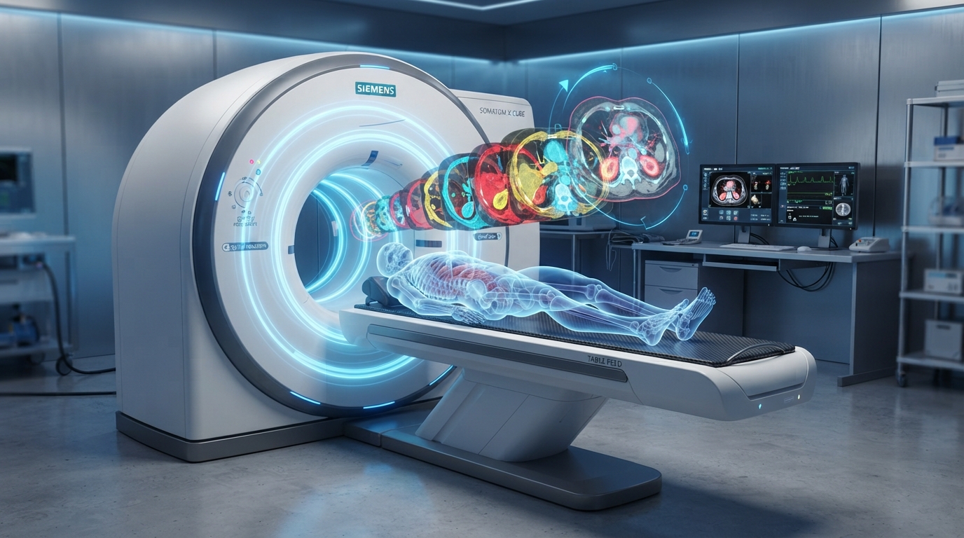
This remarkable advancement is possible because of SR CT, commonly understood as Spiral (or Helical) Computed Tomography. Unlike earlier generations of CT technology that captured images one slice at a time, spiral CT continuously rotates around the patient while the examination table moves smoothly through the scanner. The result is faster image acquisition, improved detail, and more accurate diagnosis across a wide range of medical conditions.
From emergency medicine and cancer detection to cardiovascular imaging and trauma care, SR CT has become one of the most valuable diagnostic tools in modern healthcare. Understanding how it works and why it matters offers insight into the technological innovations shaping today’s medical practice.
What Is SR CT?
SR CT, or Spiral Computed Tomography, is an advanced form of CT imaging that creates detailed cross-sectional images of the body’s internal structures using X-rays and sophisticated computer processing.
The defining feature of spiral CT is its continuous scanning motion. As the X-ray tube rotates around the patient, the examination table moves steadily through the scanner. This movement creates a spiral or helical path, allowing the machine to collect uninterrupted imaging data.
Powerful computer algorithms reconstruct this information into highly detailed images that physicians can examine from multiple angles. In many cases, these images can also be converted into realistic three-dimensional models, helping specialists visualize anatomy with exceptional clarity.
This continuous scanning process distinguishes SR CT from earlier CT systems, which relied on stopping and starting between individual image slices.
How SR CT Works
The operation of SR CT combines precision engineering with advanced computing.
When a patient lies on the scanning table, the table moves slowly through the circular opening of the scanner, known as the gantry. During this movement, the X-ray tube rotates continuously around the body while detectors positioned opposite the tube measure how much radiation passes through different tissues.
Dense materials such as bone absorb more X-rays, while soft tissues absorb less. The computer processes these differences to generate detailed cross-sectional images.
Because the scanner collects data continuously rather than in separate steps, the examination is completed more quickly while maintaining excellent image quality. This speed also reduces the chance of motion-related image blur, particularly in patients who may struggle to remain perfectly still.
Why SR CT Is Different from Conventional CT
Although both conventional CT and spiral CT rely on X-rays to create internal images, their scanning methods differ significantly.
| Feature | Conventional CT | SR CT (Spiral CT) |
|---|---|---|
| Image acquisition | Individual slices | Continuous spiral scanning |
| Scan speed | Slower | Faster |
| Motion artifacts | More common | Reduced |
| 3D image reconstruction | Limited | Excellent |
| Emergency use | Effective | Highly effective |
| Diagnostic accuracy | High | Often higher due to continuous data collection |
These differences have made SR CT the preferred technology in many hospitals, particularly where rapid diagnosis is essential.
Clinical Applications of SR CT
The versatility of SR CT explains why it is used in nearly every major medical specialty.
In emergency medicine, rapid whole-body imaging allows physicians to identify life-threatening injuries after motor vehicle accidents, falls, or other traumatic events. Internal bleeding, organ damage, fractures, and spinal injuries can often be diagnosed within minutes.
In oncology, spiral CT plays an essential role in detecting tumors, determining cancer stage, guiding biopsies, planning treatment, and monitoring therapeutic response. Its ability to capture highly detailed images helps physicians evaluate even small abnormalities that may require further investigation.
Cardiology has also benefited enormously from SR CT. Modern scanners can produce detailed images of the coronary arteries, enabling physicians to detect narrowing, plaque buildup, and certain congenital heart abnormalities without immediately resorting to invasive procedures.
Pulmonary medicine frequently relies on spiral CT to evaluate suspected pulmonary embolism, pneumonia, chronic lung disease, and lung cancer. High-resolution imaging allows specialists to examine delicate lung structures in exceptional detail.
Neurology, orthopedics, abdominal imaging, and vascular medicine similarly depend on SR CT for accurate diagnosis and treatment planning.
Advantages
The widespread adoption of SR CT reflects the many advantages it offers both patients and healthcare providers.
One of its greatest strengths is speed. Entire regions of the body can often be scanned within seconds, making the technology particularly valuable during medical emergencies.
Image quality is another major benefit. Continuous data collection produces thin image slices that can be reconstructed into detailed three-dimensional views. Surgeons, radiologists, and other specialists often rely on these reconstructions when planning complex procedures.
Reduced motion artifacts further improve diagnostic accuracy. Since scans are completed rapidly, patients spend less time holding their breath or remaining perfectly still, resulting in clearer images.
Patient comfort also improves because shorter examination times reduce anxiety and physical discomfort, especially for individuals experiencing pain or severe illness.
Limitations and Risks
Despite its many strengths, SR CT is not without limitations.
The most significant consideration is radiation exposure. Although modern scanners are designed to minimize radiation doses, CT imaging still involves higher radiation levels than standard X-rays. Physicians carefully weigh the benefits of obtaining diagnostic information against the potential risks before recommending a scan.
Some examinations also require intravenous contrast material to improve visualization of blood vessels and internal organs. While generally safe, contrast agents may occasionally trigger allergic reactions or temporarily affect kidney function in susceptible individuals.
Cost can represent another limitation. Advanced CT equipment is expensive to purchase, maintain, and operate, making access more challenging in certain healthcare settings.
Finally, while SR CT provides exceptional anatomical detail, it cannot answer every diagnostic question. Magnetic resonance imaging (MRI), ultrasound, nuclear medicine, and other imaging techniques continue to play important complementary roles depending on the clinical situation.
Preparing for an SR CT Scan
Preparation depends largely on the specific examination being performed.
Some scans require no preparation at all, allowing patients to undergo imaging immediately upon arrival.
If contrast material is needed, patients may be asked to avoid eating for several hours beforehand. Healthcare providers also review medical history, allergies, medications, and kidney function to ensure contrast can be administered safely.
During the examination, patients typically lie comfortably on the scanning table while following simple breathing instructions. Most scans are completed quickly, and patients can usually return to normal activities shortly afterward.
The procedure itself is painless, although some individuals notice temporary warmth or a metallic taste if contrast material is injected.
The Role of Artificial Intelligence
One of the most exciting developments in medical imaging is the integration of artificial intelligence into CT interpretation.
AI-assisted software can help radiologists identify subtle abnormalities, prioritize urgent findings, and improve workflow efficiency. While these systems do not replace physician expertise, they provide valuable decision-support tools that may enhance diagnostic accuracy.
Machine learning algorithms are also being developed to reduce image noise, improve reconstruction quality, and further decrease radiation exposure without compromising diagnostic performance.
As these technologies mature, the combination of SR CT and artificial intelligence promises even greater precision in disease detection and patient care.
Why Matters in Modern Healthcare
Healthcare increasingly depends on rapid, accurate decision-making.
Whether evaluating stroke symptoms, diagnosing appendicitis, detecting cancer, or assessing traumatic injuries, physicians require reliable imaging that delivers comprehensive information quickly.
SR CT fulfills this need by combining speed, precision, and versatility in a single diagnostic tool. It enables earlier diagnosis, supports minimally invasive treatments, reduces unnecessary exploratory procedures, and improves communication among multidisciplinary healthcare teams.
Its value extends beyond hospitals as well. Researchers continue using spiral CT technology to advance medical science, improve surgical planning, and develop personalized treatment strategies that enhance patient outcomes.
The Future of Spiral CT
Innovation continues to reshape CT imaging.
New scanner generations offer greater detector coverage, faster rotation speeds, lower radiation doses, and improved image resolution. Photon-counting CT, enhanced detector technology, and advanced reconstruction algorithms promise even more detailed imaging while further reducing patient exposure.
Researchers are also exploring ways to combine anatomical imaging with functional information, creating more comprehensive diagnostic tools capable of identifying disease earlier than ever before.
As medicine continues moving toward precision healthcare, SR CT will remain a cornerstone of diagnostic imaging, helping clinicians deliver faster, safer, and more individualized care.
Conclusion
SR CT has fundamentally transformed diagnostic medicine by making high-quality internal imaging faster, more accurate, and more accessible. Its continuous spiral scanning technique provides detailed anatomical information that supports physicians across virtually every medical specialty, from emergency medicine and oncology to cardiology and surgery.
While careful consideration of radiation exposure and appropriate clinical use remains essential, the benefits of spiral CT are undeniable. Faster diagnoses, improved treatment planning, and better patient outcomes have made it one of the defining technologies of modern healthcare.
As artificial intelligence, advanced detector systems, and next-generation imaging innovations continue to evolve, SR CT will likely become even more powerful, reinforcing its role as an indispensable tool in the future of medicine.